Archived Content
Information identified as archived on the Web is for reference, research or recordkeeping purposes. It has not been altered or updated after the date of archiving. Web pages that are archived on the Web are not subject to the Government of Canada Web Standards. As per the Communications Policy of the Government of Canada, you can request alternate formats on the Contact Us page.
Black Stain Root Disease
Leptographium wageneri (Kendrick) M. J. Wingfield
(= Verticicladiella wageneri (Kendrick))
(teleomorph=Ophiostoma wageneri (Goheen & Cobb) Harrington)
Deuteromycotina, Stilbellales, Stilbellaceae
Hosts:Leptographium wageneri has been reported in B.C. on Douglas-fir, lodgepole pine, western white pine, Engelmann and white spruce, and western hemlock. In other parts of North America it has also been found on mountain hemlock, ponderosa pine, and grand and white fir. In B.C., two varieties of the fungus are recognized based on host associations: Leptographium wageneri var ponderosum on pines and spruce, and Leptographium wageneri var pseudotsugae on Douglas-fir.
Distribution: Since the first report of black stain root disease in B.C. in 1976, this problem has been reported from many areas in the southern interior and coastal forests.
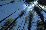
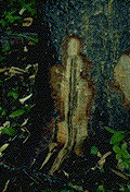
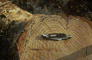
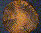
Identification: Crown symptoms in trees infected by L. wageneri are difficult to distinguish from those caused by other root diseases. Reduced leader and branch growth usually occurs quickly accompanied by foliage discoloration and thinning (Fig. 4a). Mortality occurs in "centers" similar to other root diseases. Usually by the time crown symptoms become evident, a purple-brown to black stain can be found in the main lateral roots and the base of the stem. In the stem, the stain occurs as long, tapered streaks (Fig. 4b); in cross-section it appears as narrow bands following the spring wood growth rings (Fig. 4c). Stained sapwood may be resin-soaked.
Microscopic Characteristics: Conidiophores upright, dark coloured, the upper portion with penicilliate branches; conidia hyaline, obovoid, elliptic, or clavate, base usually truncate, 2-8 x 1.7-3.8 µm, held together in a white mucilaginous mass that darkens with age.
Damage: In the interior, up to 50% of the trees have been killed in some lodgepole pine stands 60-110 years of age; infection of Douglas-fir and spruce is less common. On the coast the disease has been reported most frequently in young (15-30 years) stands of Douglas-fir although trees 60 years of age have been killed; infection of western hemlock is rare.
Remarks: Black stain is a vascular wilt; the fungus grows through infected roots in the tracheids preventing water conduction. On reaching the root collar, the fungus grows into uninfected roots and may extend three or more metres up the stem. Local spread occurs through root grafts and infection of feeder roots. Long distance spread has been attributed to root-feeding insects. Black stain root disease may make infected trees attractive to secondary bark beetles such as Ips and Pseudohylesinus spp. In addition, the fungus may be vectored by root feeding beetles into trees already weakened by Phellinus weirii and Armillaria ostoyae. In dead trees, the purple-brown to black stain may be obscured by blue stain fungi or sap rots. Blue-stain will appear in a wedge-shaped pattern (Fig. 4d), rather than in bands following annual rings (Fig. 4c). The lower bole and roots of several trees with crown symptoms in an infection center should be examined to determine the cause of the problem. Atropellis piniphila also produces a black stain, but is found in stem cankers rather than in roots.
References:
Hunt, R. S. and D. J. Morrison. 1980. Black stain root disease in British Columbia. Can. For. Serv., Forest Pest Leaf. No. 67. Victoria, B.C.
Harrington, T. C. and F. W. Cobb. 1988. Leptographium root diseases on conifers. APS Press, St. Paul, MN.
Figures
Click on any image to see the full size version.
Press "Back" on your browser to return to this screen.

Figure 4a: Crown thinning of infected lodgepole pine trees.
 Figure 4b: Black stain visible as longitudinal streaks in Douglas-fir.
 Figure 4c: Cross-section of black stain, which develops in a pattern that follows the annual rings.
 Figure 4d: In contrast, blue stain forms radially in a wedge-like pattern.
 This Web page has been archived on the Web.