Archived Content
Information identified as archived on the Web is for reference, research or recordkeeping purposes. It has not been altered or updated after the date of archiving. Web pages that are archived on the Web are not subject to the Government of Canada Web Standards. As per the Communications Policy of the Government of Canada, you can request alternate formats on the Contact Us page.
Brown Stringy Trunk Rot
Echinodontium tinctorium (Ellis & Everh.) Ellis & Everh.
(= Fomes tinctorius Ellis & Everh.)
Basidiomycotina, Aphyllophorales, Hydnaceae
Hosts: In B.C., Echinodontium tinctorium has been reported on mountain and western hemlock, amabilis, grand and subalpine fir, white and Sitka spruce, Douglas-fir, and western redcedar. In other parts of North America it has also been found on larch, Engelmann spruce, and pine True firs are highly susceptible throughout their range, western hemlock is moderately to severely attacked in specific habitats, but Douglas-fir and spruce are seldom attacked. The reports of the fungus on pine and cedar are questionable.
Distribution: Throughout host range in B.C., at high elevations in coastal forests, not reported on the Queen Charlotte Islands; restricted to western North America.
Identification: Sporophores form on living trees, generally in association with branch stubs (Figs. 9a, 9b), and may be up to 30 cm in width. The upper surface of the perennial, hoof-shaped fruiting body is hard, fissured, and generally black. The lower surface bears downward-directed spines, or teeth. These are grey to light-brown when young, turning black with age. The context of the sporophore is brick-red.
The early stage of decay appears as a light brown or water-soaked stain in the heartwood (Fig. 9c). Later the wood darkens to red-brown or yellow-brown. Small rust-coloured flecks and occasionally streaks and white channels, resembling insect tunnels, may develop. Delamination may occur along annual rings (Fig. 9d). Heartwood in advanced decay is reduced to a brown, fibrous, stringy mass which may disintegrate forming a cavity in the tree (Fig. 9e).
Microscopic Characteristics: Hyphae in the context of the fruiting body with clamps, basidiospores smooth-echinulate, hyaline, amyloid, 5.5-8 x 3.5-6 µm. Growth in culture slow, mat white to buff, reverse brown, laccase positive, hyphae with clamps, ellipsoid chlamydospores and clavate cystidia common, Stalpers: 1 2 3 (9) (10) (11) (12) (13) (17) 18 21 (22) (23) 25 30 31 (34) 3(6) 38 39 42 44 (45) 46 48 52 53 (60) 67 (72) 80 (82) 83 85 (88) 90.
Damage: This fungus is the main cause of heart rot and volume loss in mature hemlock and true firs. Sporophores are reliable indicators of defect and are associated with substantial volumes of decay. One fruiting body usually indicates that the entire cross section of the log is decayed for a distance of 2 m above, and 2.5 m below the conk. (Fig. 9f) Decay may also be present in trees that do not bear sporophores.
Remarks: Advanced stages of decay closely resemble equivalent stages of rot associated with Stereum sanguinolentum. The common name for Echinodontium tinctorium, "Indian paint fungus," is derived from the native Indian use of the ground sporophores in the preparation of red paint pigments. Losses may be reduced by harvesting at pathological rotation age. It has also been suggested that infection might be reduced by inducing natural self-pruning of suppressed branchlets, which are considered to be the major infection courts.
References:
Malloy, O. C. 1991. Review of Echinodontium tinctorium Ell. & Ev. [1895-1990]: The Indian paint fungus. Wash. St. Univ. Coop. Ext. Serv. EB1592.
Thomas, G. P. 1958. The occurrence of the Indian paint fungus, Echinodontium tinctorium, in British Columbia. Studies in For. Path. XVIII. Can. Dept. Ag. Publ. 1041.
Figures
Click on any image to see the full size version.
Press "Back" on your browser to return to this screen.
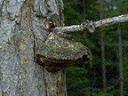
Figure 9a: Echinodontium tinctorium sporophore associated with a dead branch.
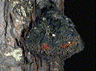 Figure 9b: Sporophore with characteristic toothed lower surface. Note the red context colour where the outer surface is chipped away.
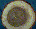 Figure 9c: Cross-section of early decay symptoms in western hemlock.
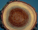 Figure 9d: Cross-section of advanced decay symptoms.
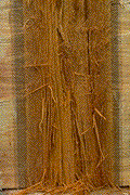 Figure 9e: Longitudinal section of advanced decay showing typical stringy brown rot symptoms.
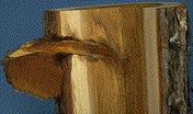 Figure 9f: Cross-section of sporophore associated with a branch and advanced heart rot.
 This Web page has been archived on the Web.